Glaucoma is defined as an elevation in intraocular pressure (IOP) beyond that which is compatible with the health of the eye. For convenience, the glaucoma syndrome can be divided into three major groups: (1) primary, (2) secondary, and (3) congenital. Primary glaucoma exhibits no overt abnormalities to account for the abnormal elevation of IOP. In secondary glaucoma, often times the most complicated, an associated disease is present within the eye. The congenital glaucomas are characterized by malformations of the anterior chamber angle (goniodysgenesis).
Glaucoma is unique in that this disease eventually involves all tissues of the eye. The incidence of glaucoma is frequently higher in certain breeds (genetic) such as the Beagle, American Cocker Spaniel, Basset Hound, Chow Chow, and Siberian Husky.
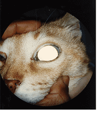
There are four hallmark clinical signs of glaucoma: a red eye; an enlarged eye; enlarged pupil; and blindness. Other clinical signs commonly observed are ocular pain and a cloudy cornea.
Diagnosis is based on the clinical history, presenting clinical signs, tonometry (which is estimation of intraocular pressure), and ophthalmoscopy. One of the most sophisticated means of determining intraocular pressure is the use of the Tonopen, which is utilized in this practice. Mormal intraocular pressure in the canine and feline is 10-25 mm /Hg. Ophthalmoscopy is necessary to evaluate the back of the eye (ocular fundus), in particular, the optic disc, in the glaucomatous dog.
Treatment of glaucoma has as its paramount purpose to lower intraocular pressure, improve patient comfort, and maintain vision (if possible). No single regime of treatment works for every patient, which is why this disease is so complex. The options to be considered are medical vs. surgical management.
Medical management options include:
-
Miotics
-
Adrenergics
-
Hyperosmotics
-
Carbonic Anhydrase Inhibitors
Most of these drugs either decrease aqueous production or increase the facility of outflow.
Surgical management options include:
-
Cyclocryotherapy – application of intense cold directly through the bulbar conjunctiva and sclera to the ciliary body to reduce the rate of aqueous humor formation.
-
Intraocular prosthesis – this is a cosmetic procedure performed on a blind, end-stage globe. The intraocular econtents are remocved and replaced with an implant. Usually, minimal long-term medical therapy is necessary.
-
Enucleation – removal of a painful, end-stage glaucomatous globe.
ATLANTA VETERINARY EYE CLINIC
33 Avondale Plaza North
Avondale Estates, GA 30002
Map & Directions | Email
OFFICE HOURS
M W F 9:00 am - 3:00 pm
T Th 9:00 am - 6:00 pm
Saturdays Closed
Sun Closed
OFFICE HOURS
CALL TO SCHEDULE AN APPOINTMENT
WE ACCEPT